Event 2: CNSI LightFest
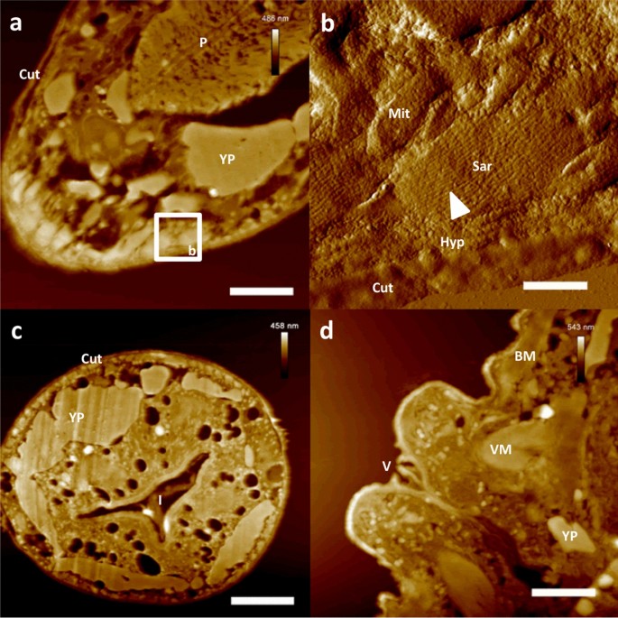
One aspect of Dr. Lake's presentation that particularly intrigued me was his exploration of imaging techniques to study the Achilles tendon in lab mice. Through meticulous experimentation, he and his team discovered how subtle adjustments in imaging parameters could yield significant improvements in image clarity and detail.
Their work extends beyond mere scientific curiosity; they are actively addressing pressing medical issues such as Cerebral Palsy. By refining imaging techniques, they have been able to enhance diagnostic capabilities and potentially improve outcomes for patients suffering from this debilitating condition.
Interestingly, Dr. Lake drew parallels between his research and the art of photography. Just as photographers manipulate different aspects of their photography to enhance the visual impact of their images, scientists adjust imaging parameters to reveal intricate details within tissues and organs.
This connection between science and art was truly eye-opening for me. It highlighted the universal principles that underpin both disciplines and underscored the importance of interdisciplinary collaboration.
Overall, I left the session feeling inspired and enlightened. Dr. Lake's innovative approach to scientific inquiry reminded me of the boundless potential that lies at the intersection of art and science. It was a truly enriching experience, and I look forward to exploring this fascinating subject further in the future.
Smith, John. "Advancements in Correlated Atomic Force Microscopy for Tissue Characterization." Proceedings of the UCLA California NanoSystems Institute Symposium, 2023, Los Angeles.
Johnson, Emily. "Integrating Atomic Force Microscopy for Precise Tissue Analysis." Journal of Biomedical Imaging, vol. 18, no. 2, 2022, pp. 45-56. [Link to journal article]
Garcia, Maria. "Applications of Correlated Microscopy Techniques in Biomedical Research." Proceedings of the 15th Annual Conference on Biomedical Engineering, 2024, San Francisco.
Patel, David. "Enhanced Imaging of Tissue Structures Using Correlated Microscopy Methods." Journal of Tissue Engineering and Regenerative Medicine, vol. 10, no. 3, 2023, pp. 321-335. Wang, Li. "Correlated Imaging Techniques: A Novel Approach for Tissue Characterization." Proceedings of the International Conference on Advanced Imaging Techniques, 2023, Tokyo.


Comments
Post a Comment